![granules inside natural killer cells](https://www.cuimc.columbia.edu/sites/default/files/styles/cola_media_200_21_9/public/media/images/2021-08/orange_lytic_granules_in_nk_cell.jpg?itok=X3YggFDk 200w, https://www.cuimc.columbia.edu/sites/default/files/styles/cola_media_260_21_9/public/media/images/2021-08/orange_lytic_granules_in_nk_cell.jpg?itok=dta5dp0R 260w, https://www.cuimc.columbia.edu/sites/default/files/styles/cola_media_320_21_9/public/media/images/2021-08/orange_lytic_granules_in_nk_cell.jpg?itok=ZIM20iF3 320w, https://www.cuimc.columbia.edu/sites/default/files/styles/cola_media_400_21_9/public/media/images/2021-08/orange_lytic_granules_in_nk_cell.jpg?itok=XQwJfkOZ 400w, https://www.cuimc.columbia.edu/sites/default/files/styles/cola_media_520_21_9/public/media/images/2021-08/orange_lytic_granules_in_nk_cell.jpg?itok=_WOAx0r- 520w, https://www.cuimc.columbia.edu/sites/default/files/styles/cola_media_640_21_9/public/media/images/2021-08/orange_lytic_granules_in_nk_cell.jpg?itok=KwM0hYts 640w, https://www.cuimc.columbia.edu/sites/default/files/styles/cola_media_800_21_9/public/media/images/2021-08/orange_lytic_granules_in_nk_cell.jpg?itok=z8AJUJpi 800w, https://www.cuimc.columbia.edu/sites/default/files/styles/cola_media_1040_21_9/public/media/images/2021-08/orange_lytic_granules_in_nk_cell.jpg?itok=l4EALqSD 1040w, https://www.cuimc.columbia.edu/sites/default/files/styles/cola_media_1280_21_9/public/media/images/2021-08/orange_lytic_granules_in_nk_cell.jpg?itok=iWkUwPVj 1280w, https://www.cuimc.columbia.edu/sites/default/files/styles/cola_media_1600_21_9/public/media/images/2021-08/orange_lytic_granules_in_nk_cell.jpg?itok=Jbv6-Rb2 1600w)
Fat ‘Shield’ Protects Immune Cells—and Some Cancers—from Destruction
A newly discovered fat "shield" that prevents natural killer cells from being destroyed by their own deadly biological weapons also allows some cancer cells to evade an immune system attack, a study at Columbia University has found.
The findings, which may lead to new treatments for aggressive cancers, were reported Aug. 3 in the journal PLoS Biology by scientists in the Department of Pediatrics at Columbia University Vagelos College of Physicians and Surgeons.
![](https://www.cuimc.columbia.edu/sites/default/files/styles/cola_media_200/public/media/images/2021-07/striking_image_2.png?itok=WK_4k3aE 200w, https://www.cuimc.columbia.edu/sites/default/files/styles/cola_media_260/public/media/images/2021-07/striking_image_2.png?itok=R3lmgoJU 260w, https://www.cuimc.columbia.edu/sites/default/files/styles/cola_media_320/public/media/images/2021-07/striking_image_2.png?itok=oMe5jLuL 320w, https://www.cuimc.columbia.edu/sites/default/files/styles/cola_media_400/public/media/images/2021-07/striking_image_2.png?itok=9z68UNKP 400w, https://www.cuimc.columbia.edu/sites/default/files/styles/cola_media_520/public/media/images/2021-07/striking_image_2.png?itok=PLZK1kTQ 520w, https://www.cuimc.columbia.edu/sites/default/files/styles/cola_media_640/public/media/images/2021-07/striking_image_2.png?itok=I0BNBuT1 640w, https://www.cuimc.columbia.edu/sites/default/files/styles/cola_media_800/public/media/images/2021-07/striking_image_2.png?itok=bUUqEXaC 800w, https://www.cuimc.columbia.edu/sites/default/files/styles/cola_media_1040/public/media/images/2021-07/striking_image_2.png?itok=CJE0yili 1040w, https://www.cuimc.columbia.edu/sites/default/files/styles/cola_media_1600/public/media/images/2021-07/striking_image_2.png?itok=uNjSuEJ3 1114w, https://www.cuimc.columbia.edu/sites/default/files/styles/cola_media_1280/public/media/images/2021-07/striking_image_2.png?itok=RJuRyl4_ 1280w)
A 3-dimensional model of a natural killer cell (purple) with granules (yellow) attaching to a target cell (gray). A pseudocolor scale shows differences in packing density of lipids on the natural killer cell membrane, with warmer colors indicating higher density. (Orange laboratory)
Natural killer cells are prolific assassins
Natural killer cells are our body’s first line of defense against pathogens and cancer cells, always present and ready to strike at a moment’s notice.
Natural killer cells are efficient assassins that can eliminate up to six infected or cancer cells each day. The deadly immune cells grab onto their target and blast it with toxic substances—proteins and enzymes—that punch holes in the cell’s membrane. These substances are not especially selective and could easily destroy the natural killer cell’s membrane during the attack.
But if these substances are so deadly, how do natural killer cells survive the blast? “I’ve been working on natural killer cells since the early 1990s, and every time I gave a talk about these cells, someone always asked that question,” says study leader and immunology expert Jordan Orange, MD, PhD, the Reuben S. Carpentier Professor of Pediatrics and chair of the Department of Pediatrics at Columbia University Vagelos College of Physicians and Surgeons. “And nobody really knew until now.”
Shielded by fat
Yu Li, a graduate student working with Orange to understand how natural killer cells work and co-author of the study, thought the answer might lie in the double layer of lipids—a type of fat—that makes up the outer membranes of all cells. Compared with other cells, Li noticed, the membranes of natural killer cells looked more orderly and more densely packed with lipids when viewed under a microscope.
“There were a lot of hypotheses about why natural killer cells don’t kill themselves during their attack on other cells, but they all proposed there might be a magic, unknown protein protecting these cells,” Li says. But Li had doubts. “Based on biophysical considerations, I didn't think a protein would be strong enough to protect the cells. When I looked at the cells, I thought of lipids.”
![false color image of cell membranes](https://www.cuimc.columbia.edu/sites/default/files/styles/cola_media_200/public/media/images/2021-08/yks_cell_compaison.jpg?itok=NL2xQTZR 200w, https://www.cuimc.columbia.edu/sites/default/files/styles/cola_media_260/public/media/images/2021-08/yks_cell_compaison.jpg?itok=nf_q6MUe 260w, https://www.cuimc.columbia.edu/sites/default/files/styles/cola_media_320/public/media/images/2021-08/yks_cell_compaison.jpg?itok=DV6IHuEL 320w, https://www.cuimc.columbia.edu/sites/default/files/styles/cola_media_400/public/media/images/2021-08/yks_cell_compaison.jpg?itok=FGmObExQ 400w, https://www.cuimc.columbia.edu/sites/default/files/styles/cola_media_520/public/media/images/2021-08/yks_cell_compaison.jpg?itok=6KV7yk5k 520w, https://www.cuimc.columbia.edu/sites/default/files/styles/cola_media_640/public/media/images/2021-08/yks_cell_compaison.jpg?itok=gE-XTKpS 640w, https://www.cuimc.columbia.edu/sites/default/files/styles/cola_media_800/public/media/images/2021-08/yks_cell_compaison.jpg?itok=m0pr8OGV 800w, https://www.cuimc.columbia.edu/sites/default/files/styles/cola_media_1040/public/media/images/2021-08/yks_cell_compaison.jpg?itok=Q_1ZwiNt 1040w, https://www.cuimc.columbia.edu/sites/default/files/styles/cola_media_1280/public/media/images/2021-08/yks_cell_compaison.jpg?itok=V5EcFEF- 1280w, https://www.cuimc.columbia.edu/sites/default/files/styles/cola_media_1600/public/media/images/2021-08/yks_cell_compaison.jpg?itok=lYcuUWMu 1600w)
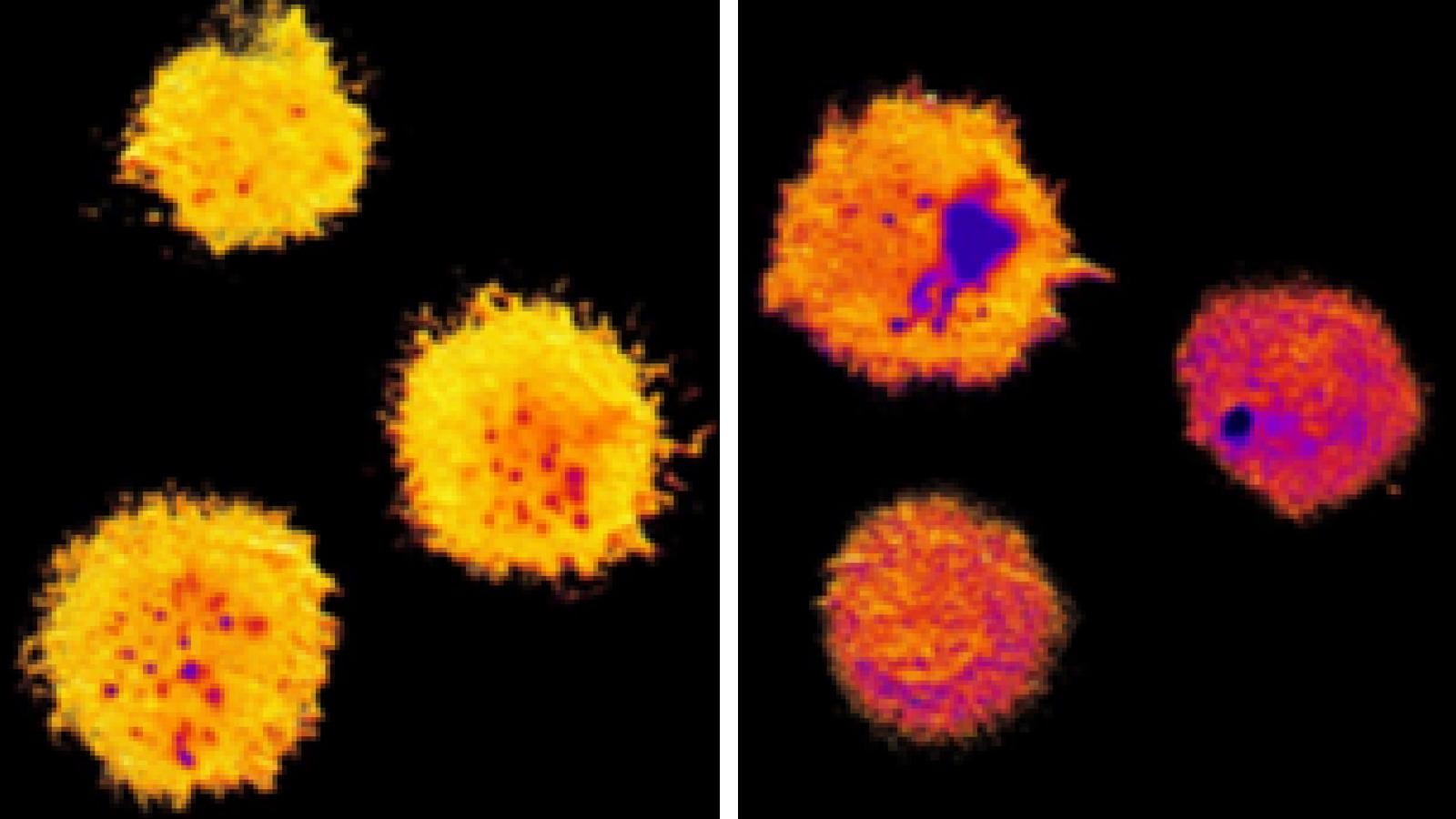
Natural killer cells with more fat packed into their membranes (as represented by the yellow color) are protected from the toxic substances deployed when the cell attacks. (Orange laboratory)
Li put his theory to the test: He exposed the membranes to a compound that weakens the structure of the lipid layer. With less dense and less orderly membranes, the natural killer cells were unprotected from their own toxic blast—and perished along with their targets.
Reinforcement arrives before natural killer cells attack
To ensure their ability to survive, natural killer cells reinforce their membranes immediately before they launch an attack, Li found. Small granules that contain the deadly substances move to the outer edge of the natural killer cell. As the granule releases its cargo into the space between the killer and target cells, its own unusually dense lipid membrane merges with and reinforces the natural killer cell membrane.
“In essence, Li found that the membrane turns into a blast shield,” Orange says. “And the protection comes from the way the membrane’s lipids are arranged. When the lipids are arranged in a more orderly fashion, more lipids can be packed into the membrane. The toxic substances simply can’t find a way into the membrane.”
![granules inside natural killer cells](https://www.cuimc.columbia.edu/sites/default/files/styles/cola_media_200/public/media/images/2021-08/orange_lytic_granules_in_nk_cell.jpg?itok=fJy8S3CF 200w, https://www.cuimc.columbia.edu/sites/default/files/styles/cola_media_260/public/media/images/2021-08/orange_lytic_granules_in_nk_cell.jpg?itok=IMYFWB5F 260w, https://www.cuimc.columbia.edu/sites/default/files/styles/cola_media_320/public/media/images/2021-08/orange_lytic_granules_in_nk_cell.jpg?itok=EvDBAA3q 320w, https://www.cuimc.columbia.edu/sites/default/files/styles/cola_media_400/public/media/images/2021-08/orange_lytic_granules_in_nk_cell.jpg?itok=cLutlGpP 400w, https://www.cuimc.columbia.edu/sites/default/files/styles/cola_media_520/public/media/images/2021-08/orange_lytic_granules_in_nk_cell.jpg?itok=z_TjG5pc 520w, https://www.cuimc.columbia.edu/sites/default/files/styles/cola_media_640/public/media/images/2021-08/orange_lytic_granules_in_nk_cell.jpg?itok=anwm7yLQ 640w, https://www.cuimc.columbia.edu/sites/default/files/styles/cola_media_800/public/media/images/2021-08/orange_lytic_granules_in_nk_cell.jpg?itok=vWWJxfVN 800w, https://www.cuimc.columbia.edu/sites/default/files/styles/cola_media_1040/public/media/images/2021-08/orange_lytic_granules_in_nk_cell.jpg?itok=s4KLqmrX 1040w, https://www.cuimc.columbia.edu/sites/default/files/styles/cola_media_1280/public/media/images/2021-08/orange_lytic_granules_in_nk_cell.jpg?itok=DF6Dlu1q 1280w, https://www.cuimc.columbia.edu/sites/default/files/styles/cola_media_1600/public/media/images/2021-08/orange_lytic_granules_in_nk_cell.jpg?itok=xR659LTL 1600w)
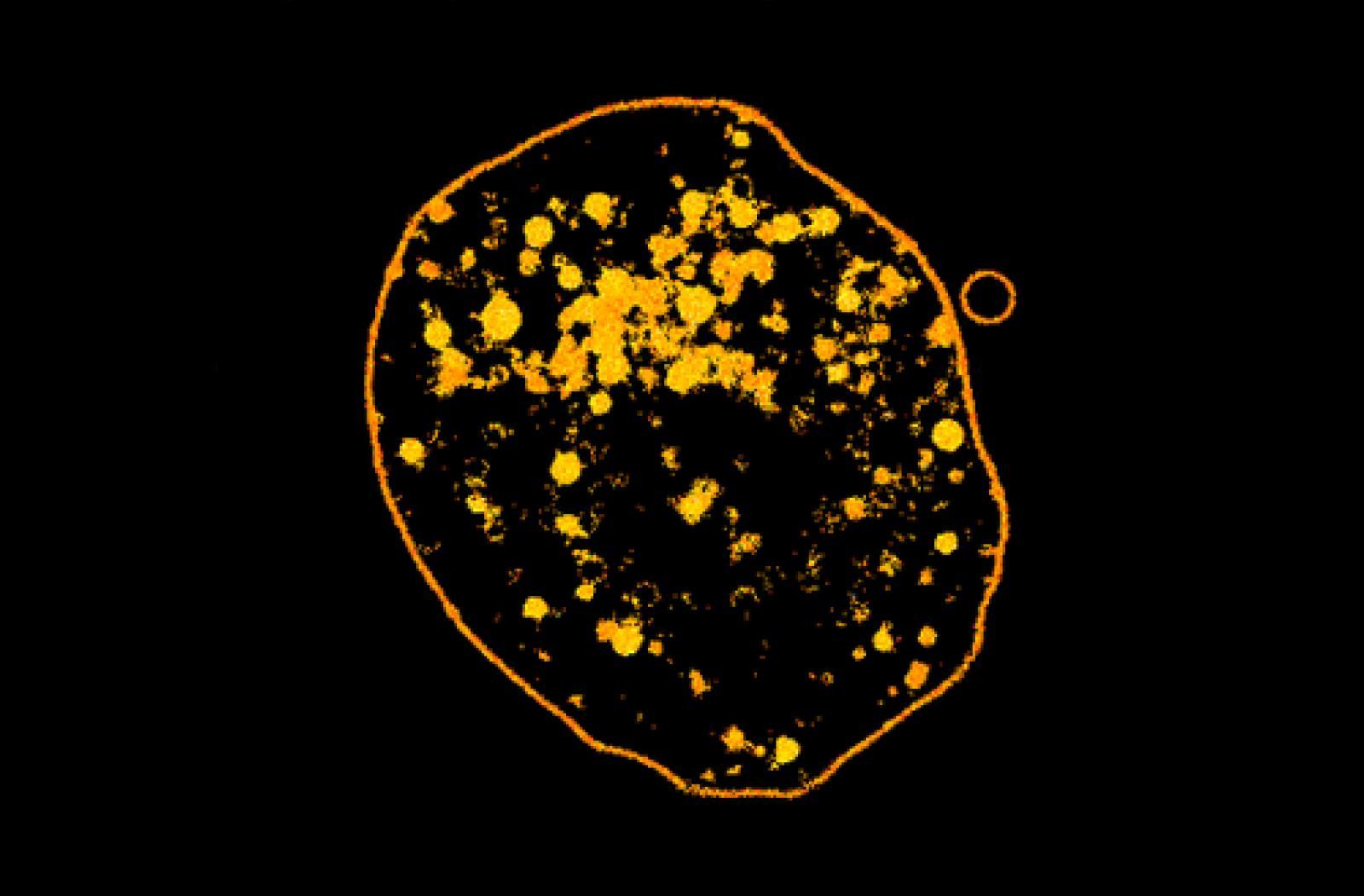
Granules (in orange) reinforce the cell membrane with extra fat to protect natural killer cells from their own weapons. (Orange laboratory)
Natural killer cells are not the only ones to adopt lipid blast shields, Li and Orange also found.
At least some cancer cells have adopted the defense to protect themselves during attacks by natural killer cells (and possibly from cytotoxic T cells, another immune cell that uses lipids for protection).
Li found that cells from an aggressive breast cancer known to be impervious to natural killer cells fortify their membranes during the attack. The fortification is essential in protecting the cancer cells, Li discovered, because when he added a membrane compound that disrupts lipid packing, the cancer cells were made vulnerable.
“We don’t know yet if this is a general mechanism by which cancer cells resist natural killer cells,” Li says. “If it is generalizable, we can start to think of therapies that disrupt the tumor cell membrane and make it more susceptible to attack by the immune system.”
References
More information
The study, titled ‘Degranulation enhances presynaptic membrane packing, which protects NK cells from perforin-mediated autolysis,” was published Aug. 3 in PLoS Biology.
The study was supported by the National Institutes of Health (R01 AI067946-14).
The authors report no financial or other conflicts of interest.